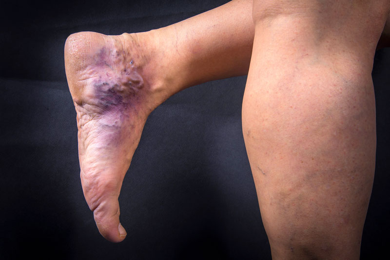
Hemosiderin staining is used to visualize blood in the tissues to determine where bleeding occurred. Hemosiderin is the main iron-containing protein found in red blood cells. It helps in staining the tissues to help us identify the injury site. Hemosiderin can be dyed in both living and fixed tissue samples.
Hemosiderin staining detects and visualizes iron in biological specimens. It is often used to determine the amount of iron stored within the body and whether there is excess iron. Hemosiderin staining is not used to diagnose or treat any disease.
Hemosiderin staining detects and visualizes iron in biological specimens. It is often used to determine the amount of iron stored within the body and whether there is excess iron. Hemosiderin staining is not used to diagnose or treat any disease. While this technique can be useful for determining the cause of death in forensic cases, it is not commonly used in medical practice.
The Hemosiderin Staining Test measures levels of free iron in the body. It is important for detecting certain conditions that involve low levels of iron in the body. A common use for this test is for screening purposes. In addition, this test can help detect diseases like thalassemia or sickle cell anemia, which are associated with lower iron levels.

What is hemosiderin staining?
Hemosiderin staining is a histological technique used to visualize iron in biological specimens. Hemosiderin staining is also used to detect and localize iron in the body. Hemosiderin is the dark pigment produced by erythrocytes when they die. It is a product of hemoglobin degradation. Hemosiderin is found in blood cells and tissues damaged or altered by infection, inflammation, or injury. Hemosiderin-containing cells can also appear in response to increased levels of circulating iron in the body.
What does it look like in your body?
To do this hemosiderin staining technique, you must first collect a blood sample from the patient. Then, you must wash the blood sample to remove red blood cells. Mixing the blood sample with a chemical solution would be best. Then, you can either use a microscope to view the blood samples under the microscope, or you can use a light microscope.
What causes hemosiderin?
Hemosiderin, or iron-laden macrophages, is formed when red blood cells die. Several reasons can lead to hemosiderin formation.
The most common reason is iron overload. This is a condition where there is too much iron in the body. It may develop due to the overuse of medications or supplements.
The second most common cause of hemosiderin is hemolysis. Hemolysis is the destruction of red blood cells. This can occur as a side effect of certain medications or diseases.
Hemolytic anemia is a rare form of anemia. It occurs when the body produces too few red blood cells. In this case, the body cannot compensate for losing red blood cells and may begin to hemolyze them.
Another cause of hemosiderin is hemorrhage. Bleeding is the loss of blood from an area of the body. When this happens, the body’s natural response is to break down blood cells.
How to Do Hemosiderin Stain Removal
First things first. Hemosiderin stains are not technically stains. They are pigments, and they are found in red blood cells. There are two types of hemoglobin in red blood cells: Hemoglobin A, which stores oxygen, and hemoglobin B, which keeps carbon dioxide.
These pigments absorb the light normally reflected off of surrounding objects, causing the red color to appear. The coloration is not permanent, and it is usually removed by washing. However, if the coloration remains long, a hematoxylin and eosin (H&E) stain will be necessary to remove it.
This is where the real work begins. The H&E stain must be done in the lab, which is time-consuming. On the other hand, Hemosiderin stains are visible under a microscope and can be used to evaluate iron levels in the body. A small tissue sample is stained and examined under a microscope to detect iron.
Hemosiderin Stain Removal – How It Works
Hemosiderin is a compound made from iron, oxygen, and nitrogen. When hemoglobin (red blood cells) die, the hemoglobin breaks down into hemosiderin. Hemosiderin can be found in both living and dead tissue. Hemosiderin is visible as a brownish-red color in tissue samples.
In living tissue, it can be seen as a reddish-purple color. In dead tissue, it appears as a gray-brown color. Hemosiderin stain removal is achieved by removing the hemosiderin from the tissue sample. Hemosiderin stain removal can be done chemically or by physical means.
Chemical methods
The first step in the chemical removal of hemosiderin is to decolorize the tissue. This is done by boiling the tissue in 10% potassium hydroxide (KOH). These are the most common methods of removing hemosiderin stains.
The next step is to oxidize the tissue. This can be done by immersing the tissue in nitric acid. This can also be done by placing the tissue in a hydrogen peroxide solution.
The last step is to remove the excess potassium ions. This is done by soaking the tissue in a 0.1M ammonium carbonate solution.
Physical methods
The first step in the physical removal of hemosiderin is to dehydrate the tissue. This is done by drying the tissue with an alcohol or acetone bath. These are less common than chemical methods but still used.
Next, the tissue is exposed to the sun. This is done by exposing the tissue to the sun for some time. The last step is to remove the excess potassium ions. This is done by soaking the tissue in a 0.1M ammonium carbonate solution.
Frequently Asked Questions Hemosiderin Staining
Q: Why is it important to determine the cause of anemia?
A: Anemia is an extremely common condition that affects many people. Many cases go unrecognized and untreated. Hemosiderin staining is a test that can help you determine what is causing the problem.
Q: What is Hemosiderin Staining?
Hemosiderin staining is a simple way to diagnose iron deficiency, which various conditions can cause.
Top Myths About Hemosiderin Staining
- Hemosiderin is a substance in red blood cells.
- Hemosiderin is the pigment that gives red blood cells their color.
- You need to prepare and stain the sample for hemosiderin staining.
Conclusion
Hemosiderin staining is an important technique for diagnosing iron deficiency. To perform a hemosiderin stain, the iron content of cells must first be released by heating the sample in nitric acid and then precipitating the iron with ammonium thiosulfate. This is called the acid-thiosulfate-iron precipitation method.
If you are new to lab work, you may not know that hemosiderin staining requires a lot of technical skill. This is because you need to prepare the sample properly. Also, it would be best to heat the piece to release the iron. Once you understand the basics of how to do hemosiderin staining, you’ll be able to apply this technique in other labs.


